
Figure 3 from MDCT signs differentiating retroperitoneal and intraperitoneal lesions diagnostic
spillage of hemorrhage into the peritoneal cavity can be picked up; a large hematoma in the retroperitoneum may be seen; may show indirect evidence of displacement of retroperitoneal structures; the presence of an abdominal aortic aneurysm with peri-aortic hemorrhage could favor a ruptured aortic aneurysm as the cause; CT.

Difference Between Intraperitoneal and Retroperitoneal Compare the Difference Between Similar
The retroperitoneum is the part of the abdominal cavity that lies between the posterior parietal peritoneum anteriorly and the posterior abdominal wall 4.. It is C-shaped on axial cross-section with convexity projecting anteriorly in the mid-line. Gross anatomy. The retroperitoneum is variably defined, mostly by the lack of consensus definition for the posterior abdominal and whether the psoas.

Anatomy of the peritoneum and peritoneal cavity Osmosis
Intraperitoneal and retroperitoneal space are the two types of cavities that occur inside the abdominal cavity, separated by the peritoneum. Furthermore, some examples of intraperitoneal organs are esophagus , stomach, jejunum , ileum , caecum, appendix, transverse and sigmoid colon, while some retroperitoneal organs include the duodenum.
Retroperitoneal Organs List
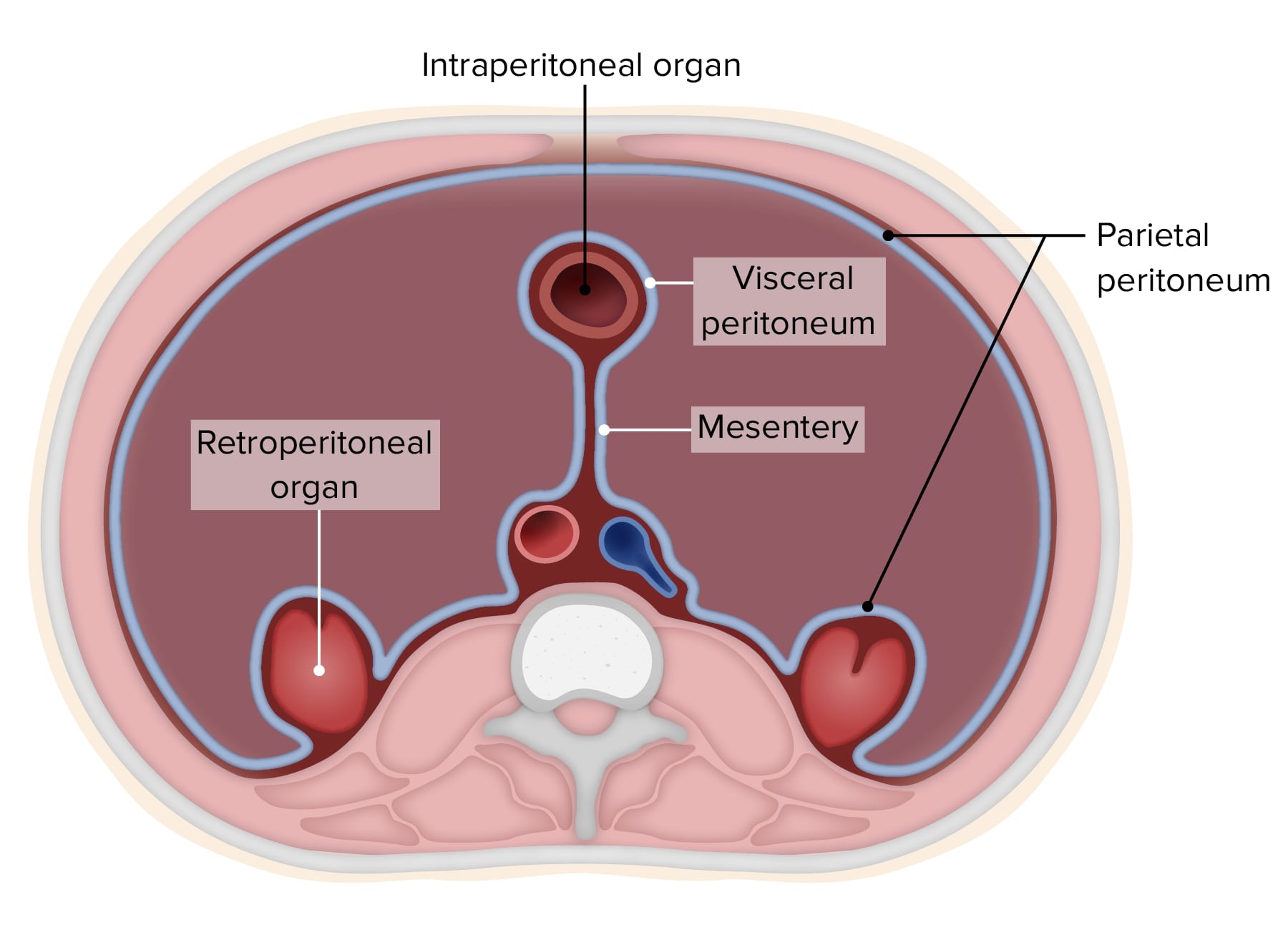
The peritoneum consists of two layers: Parietal peritoneum - an outer layer which adheres to the anterior and posterior abdominal walls.; Visceral peritoneum - an inner layer which lines the abdominal organs. It's made when parietal peritoneum reflects from the abdominal wall to the viscera.; Although in adults the peritoneum looks like it's scattered all over the place, there is a.
Peritoneum und Retroperitoneum Anatomie Lecturio
Retroperitoneal lymph nodes are located in the abdomen. They may become enlarged because of primary or secondary causes. Primary causes include things like infections or cancers that develop in the lymph nodes. Secondary causes include conditions affecting nearby organs like the kidneys or pancreas.

Peritoneal and Retroperitoneal Anatomy and Its Relevance for CrossSectional Imaging RadioGraphics
The clinical manifestations of retroperitoneal masses are nonspecific, depending on their location and relation with the adjacent structures ().The main imaging methods for the evaluation of these lesions are computed tomography (CT) and magnetic resonance imaging (MRI), imaging features facilitating the differential diagnosis, the tumor staging, and the definition of the surgical strategy, as.

Retroperitoneal Vs Intraperitoneal
Retroperitoneal hematoma is defined as bleeding into the retroperitoneal space. This clinical entity is often occult and under-recognized by clinicians and is a cause of significant morbidity and mortality. Often patients do not manifest clinically apparent signs and symptoms until a substantial amount of blood loss has occurred. It is not uncommon for patients to present in frank hemorrhagic.

Peritoneal and Retroperitoneal Anatomy and Its Relevance for CrossSectional Imaging RadioGraphics
The secondary retroperitoneal organs, which were initially intraperitoneal and became retroperitoneal structures during embryologic development due to the regression of peritoneal tissue lying on the posterior wall of the abdominal cavity (the mesentery of these structures fuse with the posterior abdominal wall), are the ascending and.
retroperitoneumillustration1 NephroPOCUS
A useful mnemonic to remember which organs are retroperitoneal is: SAD PUCKER Mnemonic S: suprarenal (adrenal) gland A: aorta/IVC D: duodenum (second, third and fourth parts) P: pancreas (except tail) U: ureters C: colon (ascending and de.

What is the Difference Between Intraperitoneal and Retroperitoneal
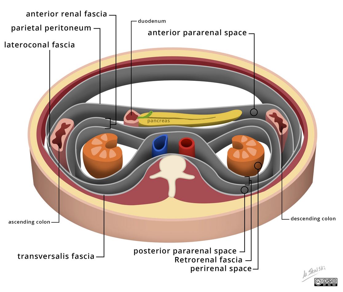
The retroperitoneum is an anatomical space located behind the abdominal or peritoneal cavity. Abdominal organs that are not suspended by the mesentery and lie between the abdominal wall and parietal peritoneum are said to lie within the retroperitoneum. Several individual spaces make up the retroperitoneum. These spaces are the anterior pararenal space, posterior pararenal space, and the.
Retroperitoneal Organs
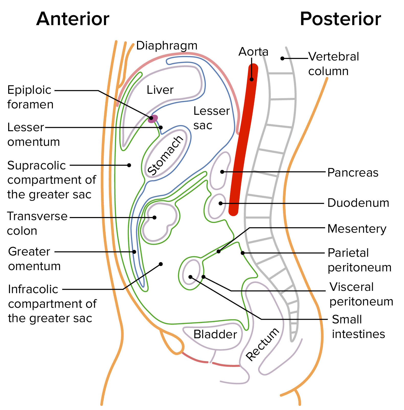
The peritoneal cavity is the space between the parietal peritoneum and the visceral peritoneum, which covers all the intraperitoneal organs (i.e., the stomach, spleen, gallbladder, liver, and part of the intestines). Approximately 50 mL of fluid is produced daily and circulates throughout the peritoneal cavity in a well defined pattern.
Intraperitoneal and Retroperitoneal Organs 3D Models, Video Tutorials & Notes AnatomyZone
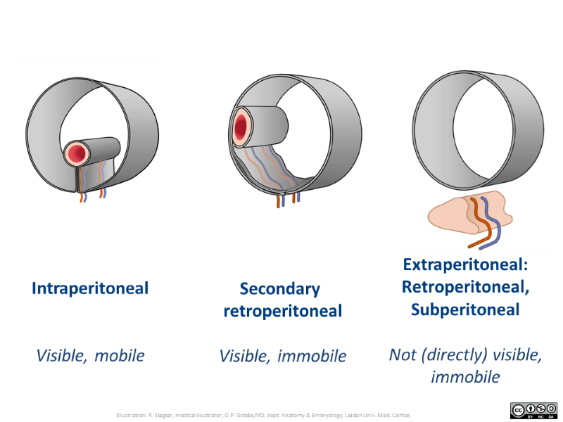
The structures in the abdomen can lie in one of three locations in relation to the peritoneum: intraperitoneal, secondary retroperitoneal or (primary) retroperitoneal. More generically, the third location should be named extraperitoneal, meaning 'outside the peritoneal cavity'.This includes, for instance, locations posterior to the peritoneal cavity, inferior to the peritoneal cavity, etc.

Intraperitoneal vs. Retroperitoneal Organs YouTube
Peritoneal Reflections. The peritoneum covers nearly all viscera within the gut and conveys neurovascular structures from the body wall to intraperitoneal viscera.. In order to adequately fulfil its functions, the peritoneum develops into a highly folded, complex structure and a number of terms are used to describe the folds and spaces that are part of the peritoneum.
Greater and lesser omentum Location, anatomy, function Kenhub
This video discusses intraperitoneal vs. retroperitoneal organs, and ends with a discussion on visceral vs. parietal peritoneum.This individual GI lecture vi.

Difference Between Intraperitoneal and Retroperitoneal Compare the Difference Between Similar
Retroperitoneal bleeding occurs when blood enters into space immediately behind the posterior reflection of the abdominal peritoneum. The organs of this space include the esophagus, aorta, inferior vena cava, kidneys, ureters, adrenals, rectum, parts of the duodenum, parts of the pancreas, and parts of the colon. Variability in presentation and etiology makes diagnosis quite difficult.[1] One.
Intraperitoneal VS Retroperitoneal organs YouTube
The structures bound by the peritoneal cavity may be intraperitoneal or retroperitoneal. The peritoneum is the serous membrane that lines the abdominal cavity. It is composed of mesothelial cells that are supported by a thin layer of fibrous tissue and is embryologically derived from the mesoderm. The peritoneum serves to support the organs of.